BAR-Net
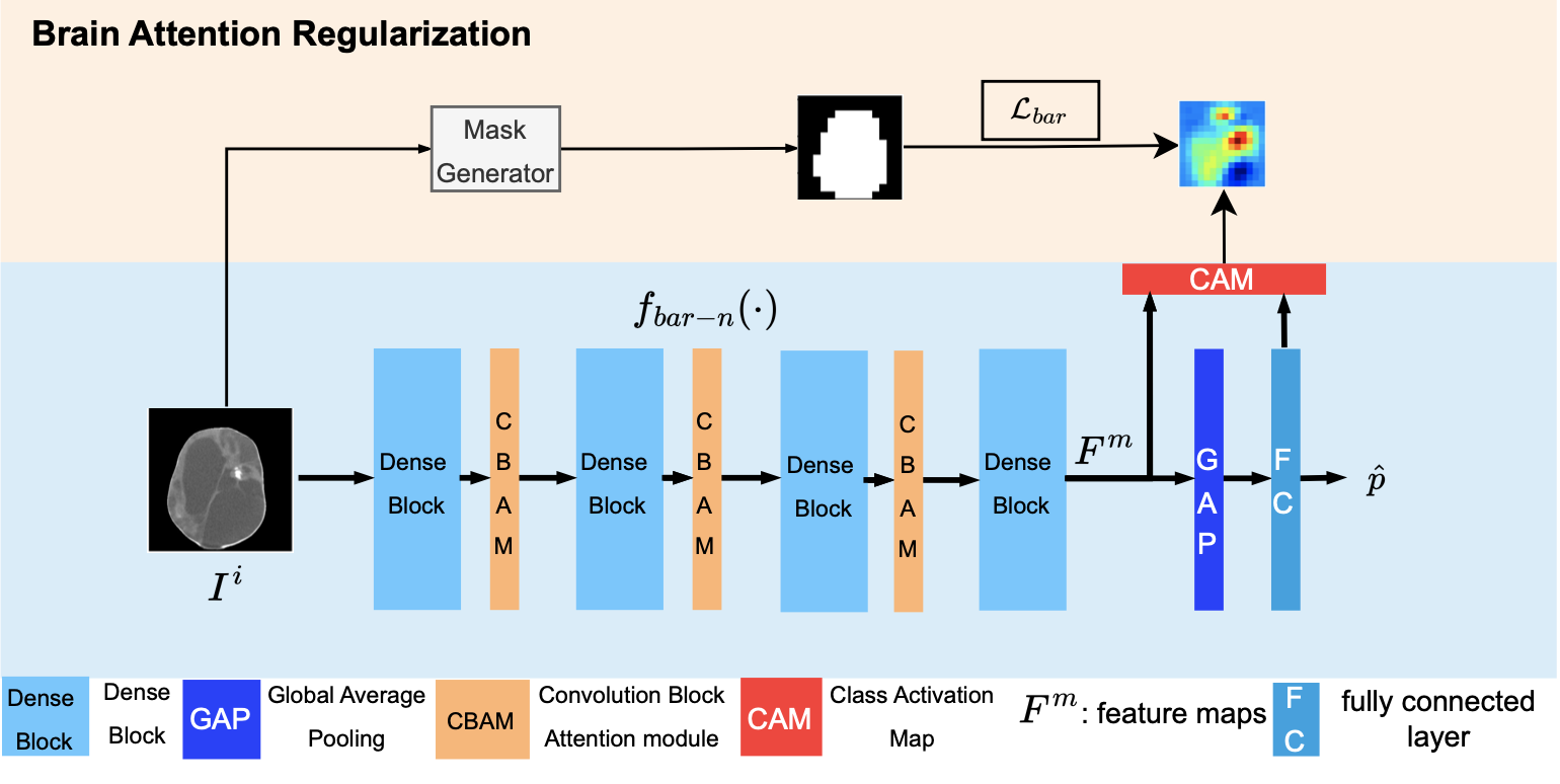
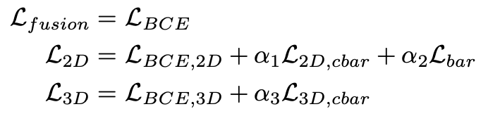
Loss function for BAR-Net:
where A is class activation map and M is brain mask.
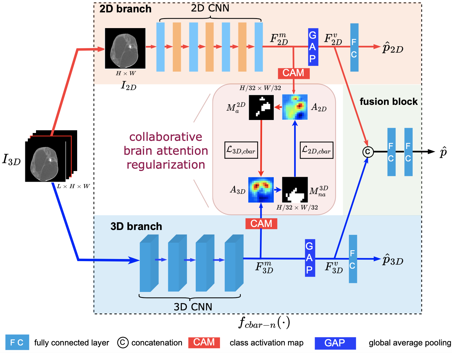
CBAR-Net
Motivation: Since the 2D branch’s input is a subset of the 3D branch’s input, it is expected that the 2D branch’s activated region is contained within the 3D branch’s activated region. Similarly, the 3D branch’s non-activated region is expected 3D to be contained within the 2D branch’s non-activated region.
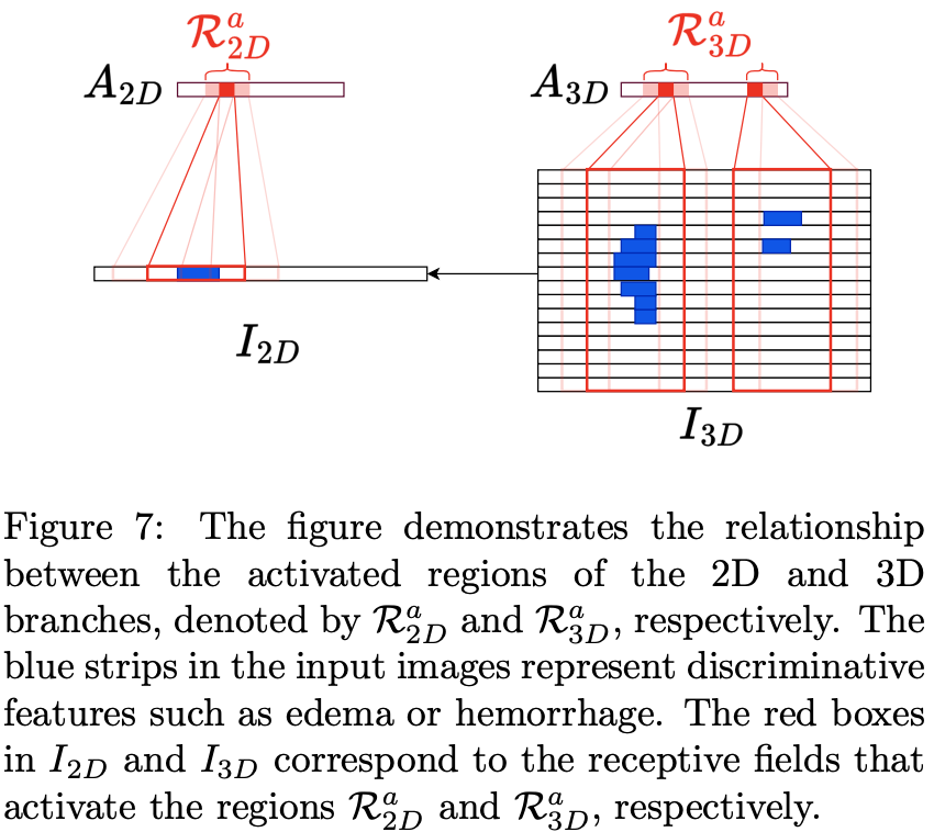
Collaborative Brain Attention Regularization To incorporate this information into the classification model, we proposed Collaborative Brain Attention Regularization and corresponding CBAR-Net.
Loss function for CBAR-Net:
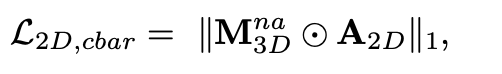
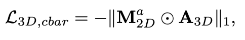
where A_2D, A_3D are class activation maps of 2D and 3D branches.
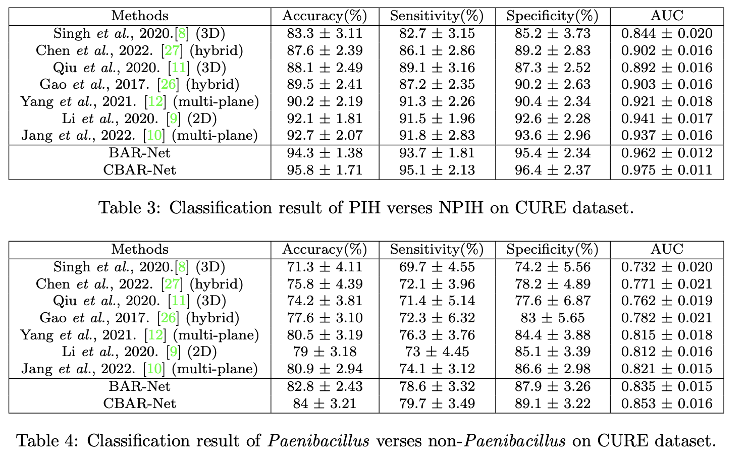
Results
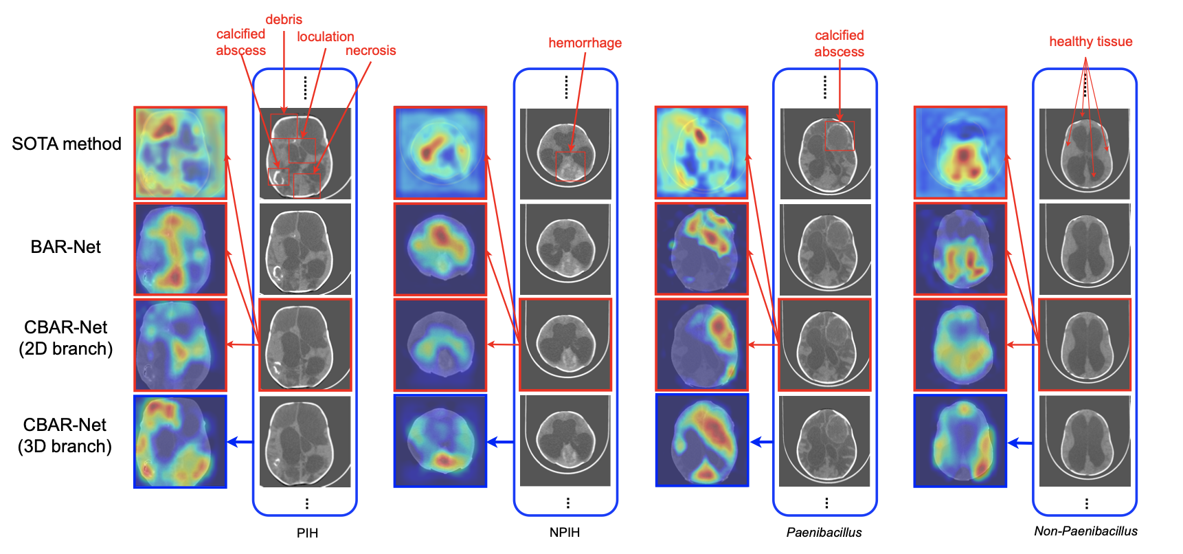
CAMs
Related Publications
Yu, Mingzhao, et al. "Infection diagnosis in hydrocephalus CT images: a domain enriched attention learning approach." Journal of Neural Engineering (2023). [JNE]