Learning Based Segmentation of CT Brain Images: Application to Post-Operative Hydrocephalic Scans

FLIS


ABSTRACT

Hydrocephalus is a medical condition in which there is an abnormal accumulation of cerebrospinal fluid (CSF) in the brain. Segmentation of brain imagery into brain tissue and CSF (before and after surgery, i.e.\ pre-op vs. post-op) plays a crucial role in evaluating surgical treatment. Segmentation of pre-op images is often a relatively straightforward problem and has been well researched. However, segmenting post-operative (post-op) computational tomographic (CT)-scans becomes more challenging due to distorted anatomy and subdural hematoma collections pressing on the brain. Most intensity and feature based segmentation methods fail to separate subdurals from brain and CSF as subdural geometry varies greatly across different patients and their intensity varies with time. We combat this problem by a learning approach that treats segmentation as supervised classification at the pixel level, i.e. a training set of CT scans with labeled pixel identities is employed. Our contributions include: 1.) a dictionary learning framework that learns class (segment) specific dictionaries that can efficiently represent test samples from the same class while poorly represent corresponding samples from other classes, 2.) quantification of associated computation and memory footprint, and 3.) a customized training and test procedure for segmenting post-op hydrocephalic CT images. Experiments performed on infant CT brain images acquired from the CURE Children extquotesingle s Hospital of Uganda reveal the success of our method against the state-of-the-art alternatives. We also demonstrate that the proposed algorithm is computationally less burdensome and exhibits a graceful degradation against number of training samples, enhancing its deployment potential.

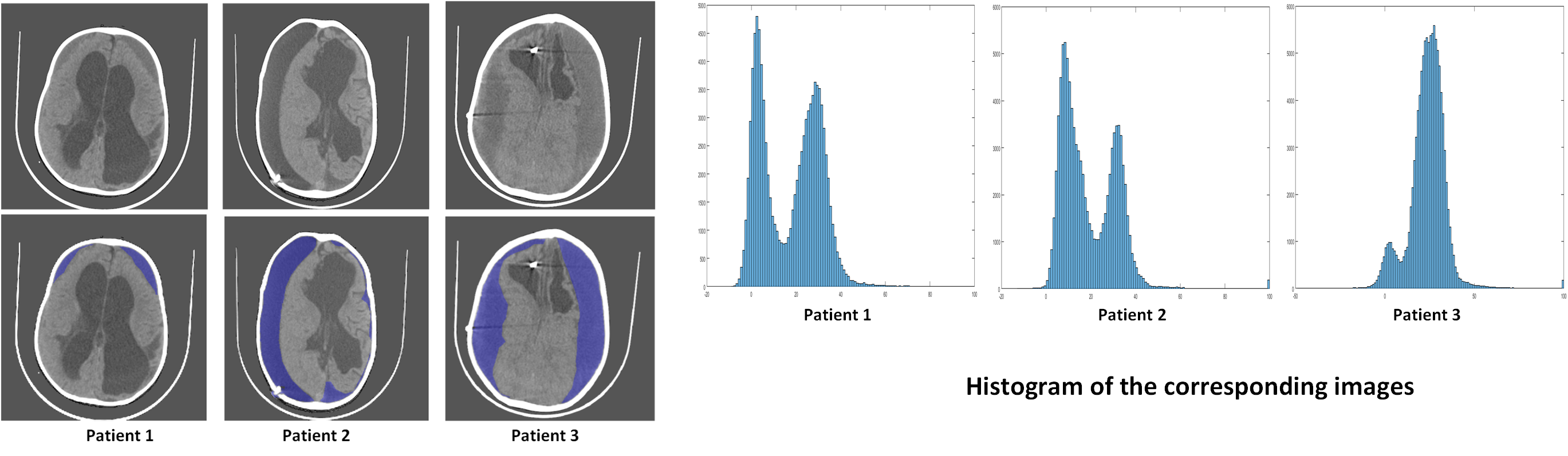
Subdural: Challenges

Idea

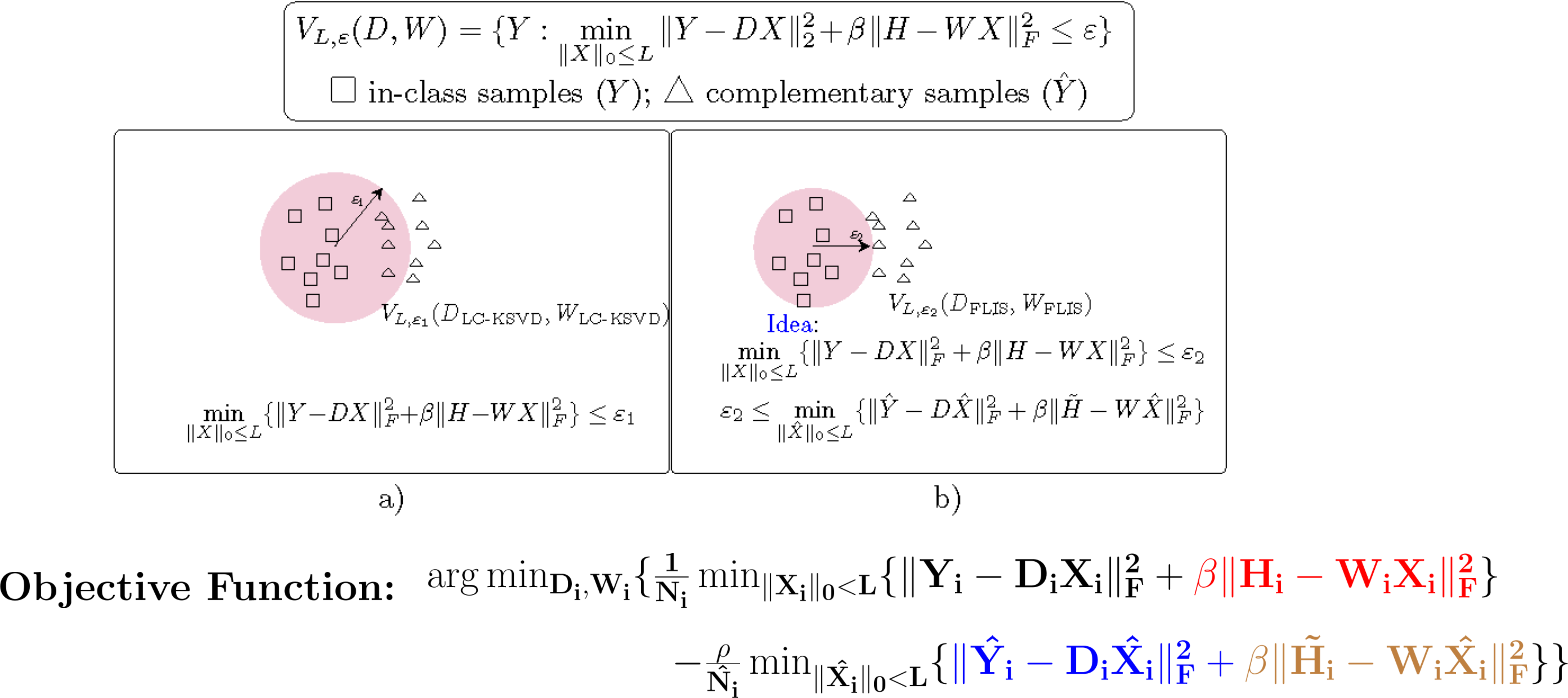
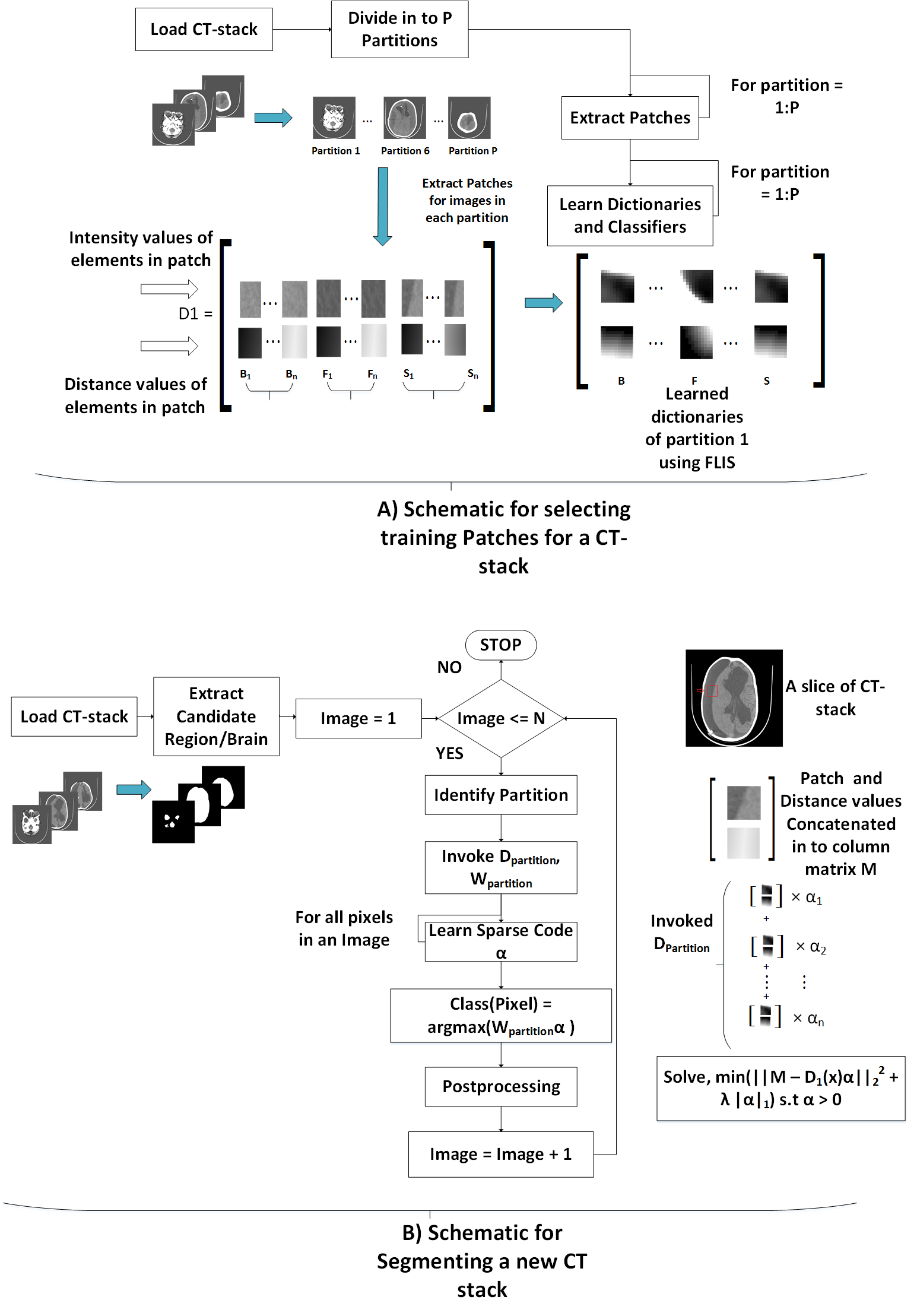
High Level Illustration

Segmentation Results

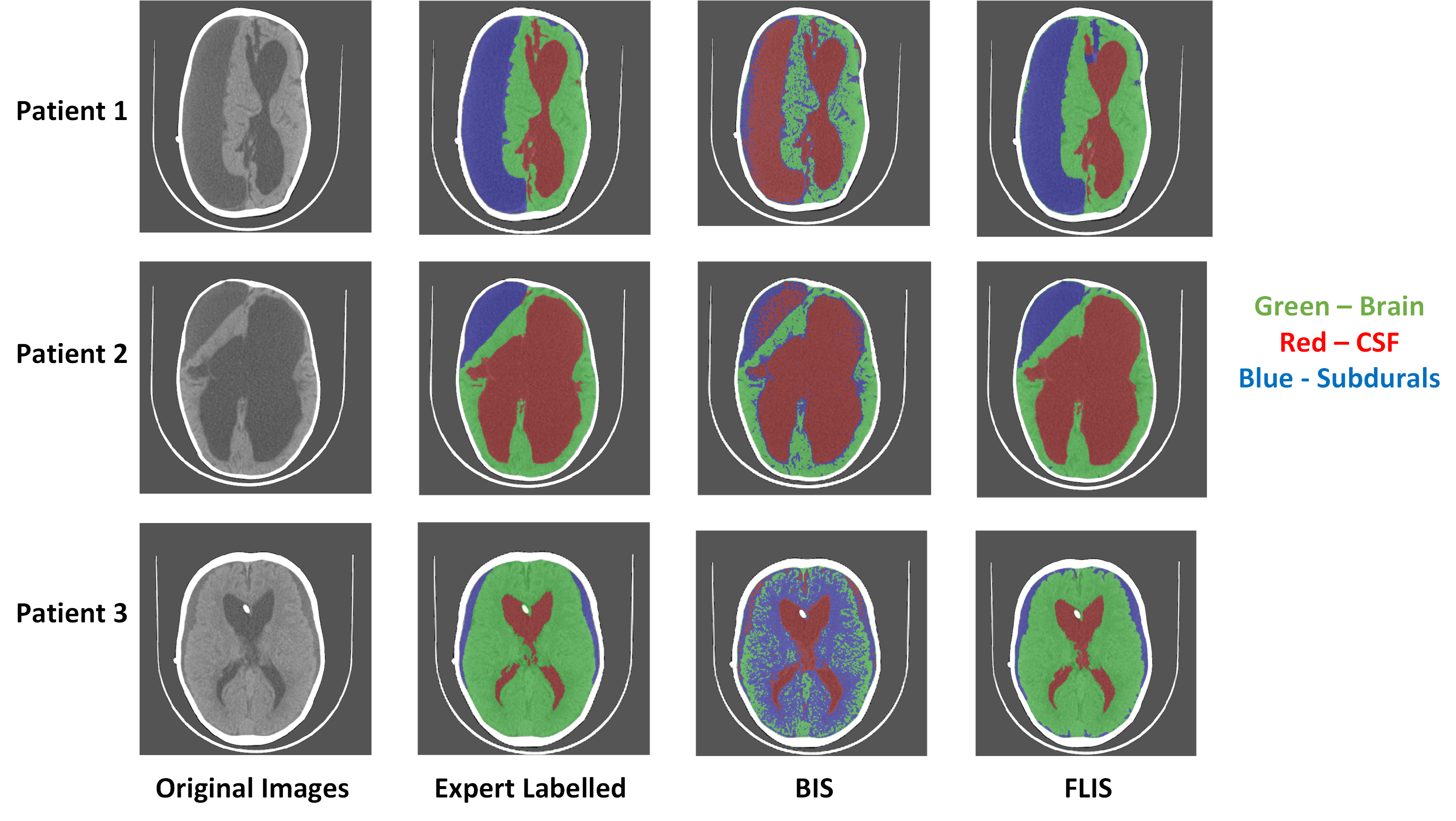
Comparison of FLIS with a traditional intensity based segmentation method.

Comparison with Deep Learning Method

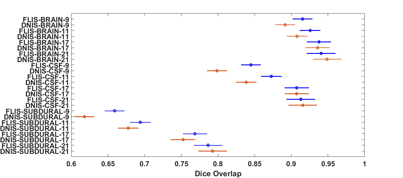
Comparison of FLIS with a deep learning method using 3-way ANOVA for different training scenarios.

Related Publications

  1. V. Cherukuri et al., "Learning Based Segmentation of CT Brain Images: Application to Postoperative Hydrocephalic Scans", IEEE Transactions on Biomedical Engineering, volume 65, issue 8, pages 1871-1884, August 2018. (IEEE Xplore)

  2. V. Cherukuri et al., "Learning based image segmentation of post-operative CT-images: A hydrocephalus case study." Neural Engineering (NER), 2017 8th International IEEE/EMBS Conference on. IEEE, 2017, Student Paper Finalist. (IEEE Xplore)

Selected References

  1. A. Makropoulos et al., “Automatic whole brain MRI segmentation of the developing neonatal brain,” IEEE Transactions on Medical Imaging, vol. 33, no. 9, 9 2014.

  2. L. Wang et al., “Integration of sparse multi-modality representation and anatomical constraint for isointense infant brain MR image segmentation,” NeuroImage, vol. 89, pp. 152–164, 2014.

  3. T. Tong et al., “Segmentation of MR images via discriminative dictionary learning and sparse coding: Application to hippocampus labeling,” NeuroImage, vol. 76, pp. 11–23, 2013.

  4. P. Moeskops et al., “Automatic segmentation of MR brain images with a convolutional neural network,” IEEE transactions on medical imaging, vol. 35, no. 5, pp. 1252–1261, 2016.

Email
ipal.psu@gmail.com

Address
104 Electrical Engineering East,
University Park, PA 16802, USA

Lab Phone:
814-863-7810
814-867-4564