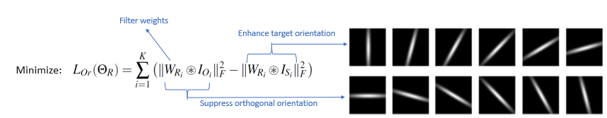
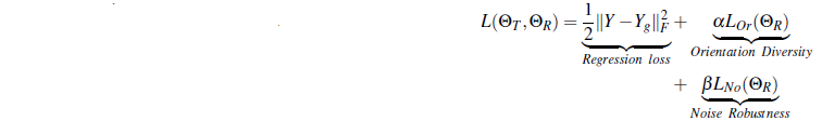
Orientation Diversity Regularizer
Representation Layer has the ability to extracts features at different orientation due to the proposed orientaion diversity regularizer.
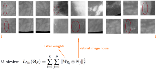
Noise Regularizer
Representation Layer supresses the curvilinear non-vessel structures due to the noise regularizer.
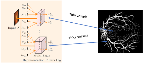
Multi-Scale Representation Layer
Representation filters trained at multiple scales to capture diversity in vessel thicknesses.
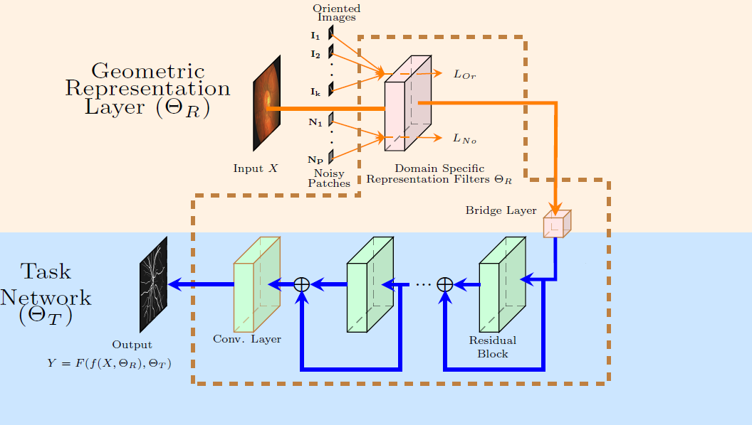
Network Structure
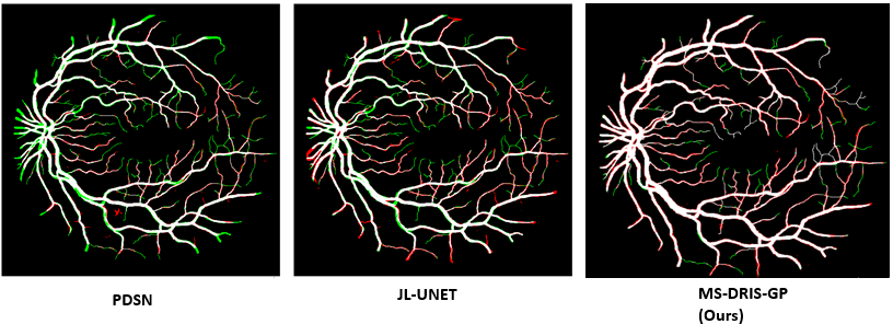
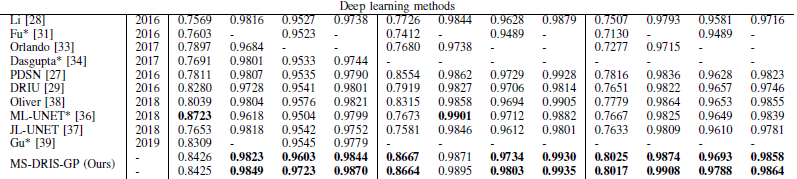
Results
Related Publications
V. Cherukuri, BG V. Kumar, R. Bala and V. Monga, "Deep Retinal Image Segmentation with Regularization Under Geometric Priors", Accepted, IEEE Trans. On Image Processing, 2019. [arXiv][IEEE Xplore]
Cherukuri, Venkateswararao, et al. "Multi-Scale Regularized Deep Network for Retinal Vessel Segmentation." 2019 IEEE International Conference on Image Processing (ICIP). IEEE, 2019. [IEEE Xplore]
Selected References
K.-K. Maninis et al., “Deep retinal image understanding,” in International Conf. on Medical Image Computing and Computer-Assisted Intervention. Springer, 2016, pp. 140–148. [paper].
Liskowski, Paweł, and Krzysztof Krawiec. "Segmenting retinal blood vessels with deep neural networks." IEEE transactions on medical imaging 35.11 (2016): 2369-2380. [paper].
Z. Yan, X. Yang, and K.-T. T. Cheng, “Joint segment-level and pixelwise losses for deep learning based retinal vessel segmentation,” IEEE Trans. on Biomedical Engineering, vol. 65, no. 9, 2018. [paper].
X. Wang, X. Jiang, and J. Ren, “Blood vessel segmentation from fundus image by a cascade classification framework,” Pattern Recognition, vol. 88, pp. 331–341, 2019. [paper].
Z. Fan, J. Lu et al., “A hierarchical image matting model for blood vessel segmentation in fundus images,” IEEE Trans. on Image Processing, vol. 28, no. 5, pp. 2367–2377, May 2019. [paper].