
Histopathological Image Data Sets
People
Please contact Prof. Vishal Monga, Department of Electrical Engineering, Pennsylvania State University, for information regarding this research.Related papers
- U. Srinivas, H. S. Mousavi, C. Jeon, V. Monga, A. Hattel, and B. Jayarao, "SHIRC: A Simultaneous sparsity model for Histopathological Image Representation and Classification", in Proc. IEEE International Symposium on Biomedical Imaging, San Francisco, CA, April 7th -11th, 2013, pp. 1106-1109.
- U. Srinivas, H. S. Mousavi, V. Monga, A. Hattel and B. Jayarao, "SHIRC: Simultaneous Sparsity Model for Histopathological Image Representation and Classification", IEEE Transactions on Medical Imaging, vol. 33, issue 5, pages 1-17, May 2014. The MATLAB code corresponding to our proposed Simultaneous Sparsity model can be downloaded here.
- Tiep H. Vu, H. S. Mousavi, V. Monga, A. U. Rao and G. Rao, "DFDL: Discriminative Feature-Oriented Dictionary Learning For Histopathological Image Classification", IEEE International Symposium on Biomedical Imaging (ISBI), Brooklyn, NY, USA, April 16th-19th, 2015.
- Tiep H. Vu, H. S. Mousavi, V. Monga, A. U. Rao and G. Rao, "Histopathological Image Classification using Discriminative Feature-Oriented Dictionary Learning", IEEE Transactions on Medical Imaging, volume 35, issue 3, pages 738-751, March 2016. The supplementary material, including but not limited to complexity analyis and MATLAB code, corresponding to our proposed Discriminative Dictionary Learning model can be found here.
Data Sets
Two different histopathological image data sets are discussed here:1. ADL Data Set
The images in this data set have been acquired by pathologists at the Animal Diagnostics Lab (ADL), Pennsylvania State University. Representative images from the data set can be downloaded here. Features of the data set:- Hematoxylin-eosin (H&E)-stained tissues
- Scanning at 40x optical magnification using a whole slide digital scanner
- Three bovine organs:
- Kidney (healthy/inflamed)
- Lung (healthy/inflamed)
- Spleen (healthy/inflamed)
- 120 images per condition per organ
- Ground truth labels for healthy and inflammatory tissue obtained via manual detection and segmentation
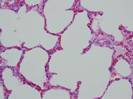 (a) Healthy lung (a) Healthy lung |
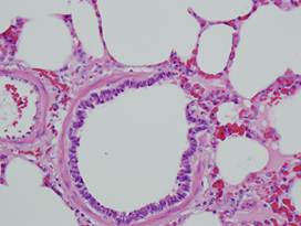 (b) Healthy lung (b) Healthy lung |
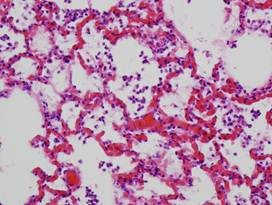 (c) Inflamed lung (c) Inflamed lung |
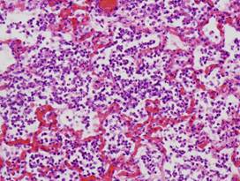 (d) Inflamed lung (d) Inflamed lung |
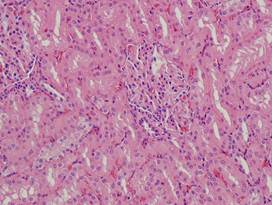 (e) Healthy kidney (e) Healthy kidney |
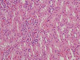 (f) Healthy kidney (f) Healthy kidney |
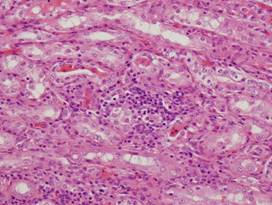 (g) Inflamed kidney (g) Inflamed kidney |
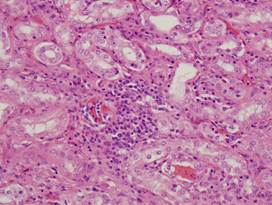 (h) Inflamed kidney (h) Inflamed kidney |
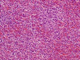 (i) Healthy spleen (i) Healthy spleen |
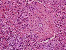 (j) Healthy spleen (j) Healthy spleen |
 (k) Inflamed spleen (k) Inflamed spleen |
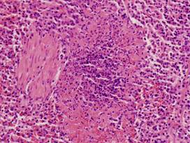 (l) Inflamed spleen (l) Inflamed spleen |
| Figure. Examples from ADL data set | |||
2. IBL Data Set
This data set contains images of human intraductal breast lesions (IBL). They have been acquired by Clarion Pathology Lab, Indianapolis and the Computer and Information Science Department, Indiana University-Purdue University Indianapolis (IUPUI), Indiana. Features of the data set:
- Two well-defined categories: usual ductal hyperplasia (UDH) and ductal carcinoma in situ (DCIS)
- Ground truth labels assigned manually by pathologists
- Total of 120 regions of interest (RoIs), or equivalently images, used for experiment: 60 training, 60 test
Images cannot be made publicly available. Please contact Prof. Murat Dundar at IUPUI for additional information.

Copyright © 2017 iPAL - All Rights Reserved